Sonography or Ultrasound imaging is used to evaluate many parts of the body. It utilizes high frequency sound waves these are not x-rays to obtain diagnostic images. 3D (4D) Sonography provides a three-dimensional view in motion and is one of the most important modern innovations in the field of Ultrasound.
The clear view of the fetus from all angles allows doctors to detect any congenital abnormalities at an early stage and chart a course for corrective measures at an early and preventable stage. The image quality is so clear and sharp that one can get a fairly accurate impression of how the baby's features will look upon birth. Sonography makes it very useful for diagnosing many parts of the body, and guiding doctors for, a range of medical conditions. Some examples of the types of sonography and what they are used to investigate include:
- Abdomen and chest – organs in the body including lungs, liver, kidneys, spleen and more
- Breasts – breast tissue, including breast lump
- Cardiac – chambers, muscle and valves of the he
- Gynaecological – female reproductive system.
- Musculoskeletal – muscles, tendons, joints
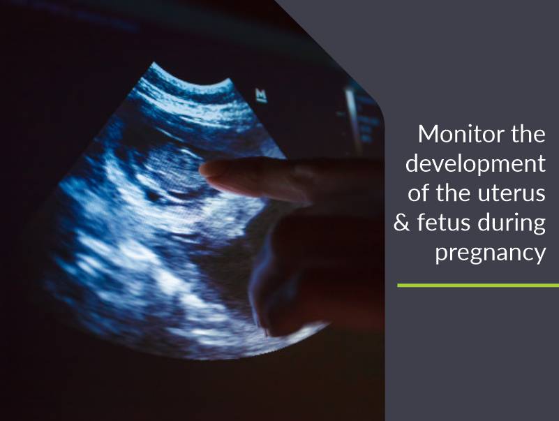
- Obstetric – fetus, cervix and placenta during pregnancy
- Small parts – male reproductive system; neck and thyroid
- Vascular – blood vessels throughout the body
WHY DO YOU NEED TO PERFORM AN ULTRASONOGRAPHY?
- Early pregnancy, including entopic pregnancy.
- Maturation of eggs and changes of endometrium in different stages of menstrual cycle.
- Development of fetuses and possible malformations of fetuses.
- Position of the fetus, position of the placenta in the uterus and changes in it. It is also possible to estimate the quantity of amniotic fluid, evaluate heart function and breathing movements of the fetus.
- Check the condition of your abdominal organs such as liver, spleen, pancreas, kidneys and gallbladder.
- For men, if they have symptoms such as enlarged organ, hernia, pancreatitis, gallstone, kidney blockage, liver cancer, appendicitis, abnormal uterine structure, abnormal fetus structure and internal tumor.
- Identify the reasons for abdominal pain and to find problems and detect damage, disease or a tumor in an organ.
- Vascular – blood vessels throughout the body
HOW IS ULTRASONOGRAPHY PERFORMED?
- Before undergoing the test, you will be provided with a comfortable gown and will be asked to remove any metal jewelry or accessory, if you are wearing any.
- The lab technician will then ask you to lie on the table, exposing your abdomen.
- A lubricant jelly will be applied on your abdomen and then with the help of a transducer, ultrasound waves of high frequency will be transmitted into your body.
- If you feel any discomfort or pain, inform the technician so that he stops the test, immediately.
- While the test is performed, pictures of the organs or the baby appear on the monitors and are captured by the technician.
- After completing the ultrasonography procedure, the technician cleans the lubricant jelly on the abdomen.
- The whole procedure takes less than 30 mins.