Benefits of 2D Echo Tests Over Traditional Cardiac Imaging Techniques

- Admin
- 09 January, 2025

Welcome to the exposition of amazing benefits of two-dimensional echocardiography tests over the old techniques of cardiac imaging. Cardiac care has completely transformed using the non-invasive method, real-time visualization as well as imaging of high resolution developed by 2D echo tests.
Here, we shall disclose why these tests are more-safe, cost effective, accessible and diagnostic in nature and lastly, they are of significance in this way as they enable close observation and supervision of cardiac health.
2D echo mean non-invasive procedures that bring safety as an alternative of cardiac catheterization which is considered invasive procedure. In the motion of catheters, 2D Echo tests use ultrasound technology to create a picture of the heart from its outside shell. This makes possible to get rid of invasive procedures risks like bleeding, infections or vessels/organs harm.
Another aspect is that 2D Echo is very non-invasive meaning very few people experience discomfort or stress during this exam. Additionally, this can be performed without incisions or perforations, which helps patients to undergo 2D ECHO tests repeatedly.
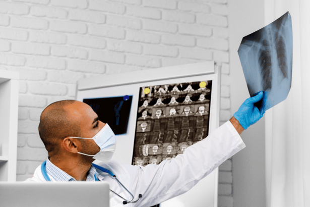
Must Read : Why X-rays Are a Vital Diagnostic Tool in Medicine
Real-time visualization of the 2D Echo scan is one of the advantages of this diagnostic method. Therefore, it allows to easily get acquainted with the structure and function of the heart. Unlike the 1D images produced by the traditional imaging tools that offer static imaging, the two-dimensional echocardiography offers real-time motion capability that is close to the actual dynamics of the cardiac motion.
Well, in-time visualization is especially significant in moments of stress echocardiography or intraoperative/operative echocardiography when there are a lot of matters for decision what doctor should make when a cardiac function is impaired.
2D Echo Tests are helpful tools that show students detailed pictures of cardiac anatomy and function in high-quality. By uing sound waves to form pictures, 2-D Echocardiography can be in the capacity for precise visualisation of cardiac structures with great resolution.
The hallmark of this specific kind of imaging is its capacity to detect with such a high level of detail. This allows for clinicians to identify abnormalities that might go unnoticed by other imaging technologies.
They permit to get detailed pictures in 2D Echo tests that are invaluable for diagnoses of multiple conditions like MI, cardiomyopathy, valvular disease, and congenital heart disease and many others.
In contrast with invasive procedures and ionizing radiation-based imaging technologies, 2D Echo test has a larger safety margin. They are no radiation exposure or use of contrast agents, therefore they have minimal risk of patients.
Therefore, these non-invasive imaging techniques are safer for everybody, even pregnant women and patients with compromised renal function or contrast agent allergies. In addition, the non-invasive attributes of 2D Echo tests makes them a preferred choice over others for routine screening and diagnostic evaluation, as there are no procedural complications involved in the process.
Compared to other cardiac imaging modalities like cardiac MRI or CT angiography, a 2D echo test is generally more cost-effective. The tools necessary for a 2D Echo examination are less expensive compared with MRI or CT scanners, and the procedure itself takes less time and fewer resources.
Furthermore, the absence of contrast agents or sedation in the 2D Echo tests lowers the expenses of medications administration and post-procedural observation. Consequently, 2D Echo checks become more available and affordable for healthcare institutions and patients who do not wish to wait for a long time to get high quality cardiovascular imaging service.
2D Echo is widely available healthcare settings around the world such as in hospitals, clinics, and outpatient imaging centers. This extensive availability will ensure that patients get timely access to cardiac imaging services regardless of their geographic location or health care resources they have.
Whether in urban locations or in remote regions, patients can undergo 2D Echo screening at facilities with ultrasound technology, ensuring the fast diagnosis and management of cardiac conditions.
The accessibility of 2D Echo tests plays a significant part in improving patient outcomes and narrowing healthcare access disparities across the diverse populations.
2D Echo tests, a versatile diagnostic tool, can evaluate many cardiac conditions from structural anomalies to functional impairments. Via employing different imaging modalities like M-mode, Doppler and color flow imaging, 2D Echo test can assess cardiac chambers, walls, valves, and blood flow patterns and obtain comprehensive diagnostic data in a single session.
2D Echo tests are characterized by their diagnostic versatility, making them appropriate in the diagnosis of various conditions like coronary artery disease, heart failure, valvular heart disease, pericardial disease, congenital heart defects, and many others.
Moreover, 2D echo tests are well-accepted by patients because little or no preparation or recovery time is needed. The procedure is performed on patients who are lying comfortably in a non-intimidating position as a transducer is moved gently over their chest area. The fact that 2D Echo tests are painless and non-invasive unlike the invasive procedures that may inflict pain and discomfort, patients can relax throughout the imaging process and cooperate.
2D echo tests are not only diagnostic but also they can be useful for following up on the disease progression and treatment response over time. 2D Echo tests performed in sequence at fixed intervals by the physicians help to monitor the changes in cardiac structure and function as well as detect early signs of disease progression or side effects associated with treatment. This longitudinal monitoring allows clinicians to be able to adapt treatment plans and interventions as necessary, resulting in better outcomes for patients and minimal adverse effects.
In summary, 2D Echo tests offer numerous advantages over traditional cardiac imaging techniques, including non-invasiveness, real-time visualization, detailed imaging, safety, cost-effectiveness, wide availability, diagnostic versatility, patient comfort, and monitoring capabilities. These benefits make 2D Echo tests a preferred choice for evaluating cardiac anatomy and function in a wide range of clinical scenarios.
Must Read : The Future of Diagnostics: Emerging Trends in Pathology Labs